

MycAwayTMPlus-Color One-Step Mycoplasma Detection Kit是利用Yeasen独特的等温扩增技术开发的针对细胞培养液中支原体污染的快速检测产品,较以往支原体检测产品本试剂盒做了升级改善,在极大程度上降低了假阳性率,提高检测的准确度并增强了阴性和阳性的辨识度。在扩增结束后,可在室温放置一段时间且不会出现由阴性转变为阳性的现象,不影响阴、阳性判断。主要原理是若细胞培养物被支原体污染,支原体DNA的保守序列会被大量、快速地扩增,使反应液由蓝紫色变成天蓝色,结果肉眼可辨,无需电泳。
太阳成集团MycAwayTMPlus-Color One-Step Mycoplasma Detection Kit可以检测多种支原体,包括细胞培养过程中常见的8种支原体。传统巢式PCR支原体法检测法易受细胞培养上清中抑制物的影响,产生假阴性结果;反应后需开盖电泳检测,增加污染导致的假阳性风险。一步法支原体检测试剂盒完全没有上述缺点,且其检测灵敏度、准确性远远高于PCR法。
1、检测种类多:可精确检测22种支原体,包括常见8种支原体;
2、结果分辨性强:检测结果参照标准清晰,检出率高,阴、阳性分辨性强;;
3、检测性能强:通过与竞品对比,试剂盒在63℃情况下从30min、40min到50min,稳定性远高于竞品,且无假阳性;
4、反应时长极限长:反应放置时长可以从60min、90min到120min均无弱阳性出现,且NTC与原始颜色无明显差别;
5、反应结束室温放置稳定性强: 63℃反应结束后,无需95℃灭活,可直接放置于室温3天,阴阳性结果与原始无明显差别。
检测性能比较
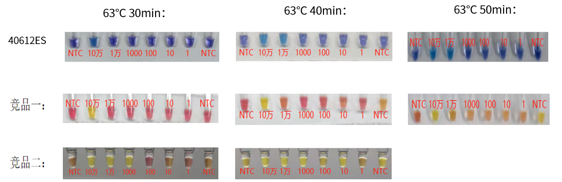
结论:
1)竞品一30min检出为10万;在40min出现NTC转阳现象;50min完全转阳;无法分辨阴性与阳性;假阳性率高。
2)竞品二30min检出为1000copies/μL,但伴随NTC出现弱阳性,且出现显色混乱,无法分辨阴性与阳性;40min完全混乱,假阳性率极高。
3)相较于竞品,40612ES 30min检出均与竞品一或竞品二相当,但其稳定性远高于竞品,无假阳性出现。
本次测试采用支原体DNA为模板,投入为1uL。
冰袋运输。-25 ~ -15℃避光保存,保质期18个月。若较长时间不用,请注意避光保存。
[1] Shi N, Yang Q, Zhang H, et al. Restoration of dystrophin expression in mice by suppressing a nonsense mutation through the incorporation of unnatural amino acids. Nat Biomed Eng. 2022;6(2):195-206. doi:10.1038/s41551-021-00774-1(IF:25.671)
[2] Zhang S, Yu F, Che A, et al. Neuroendocrine Regulation of Stress-Induced T Cell Dysfunction during Lung Cancer Immunosurveillance via the Kisspeptin/GPR54 Signaling Pathway. Adv Sci (Weinh). 2022;9(13):e2104132. doi:10.1002/advs.202104132(IF:16.806)
[3] Li Y, Xue B, Zhang M, et al. Transcription-coupled structural dynamics of topologically associating domains regulate replication origin efficiency. Genome Biol. 2021;22(1):206. Published 2021 Jul 12. doi:10.1186/s13059-021-02424-w(IF:13.583)
[4] Wu L, Xu Y, Zhao H, et al. FcγRIIB potentiates differentiation of myeloid-derived suppressor cells to mediate tumor immunoescape. Theranostics. 2022;12(2):842-858. Published 2022 Jan 1. doi:10.7150/thno.66575(IF:11.556)
[5] Tan B, Shi X, Zhang J, et al. Inhibition of Rspo-Lgr4 Facilitates Checkpoint Blockade Therapy by Switching Macrophage Polarization. Cancer Res. 2018;78(17):4929-4942. doi:10.1158/0008-5472.CAN-18-0152(IF:9.130)
[6] Yan G, Zhao H, Zhang Q, et al. A RIPK3-PGE2 Circuit Mediates Myeloid-Derived Suppressor Cell-Potentiated Colorectal Carcinogenesis. Cancer Res. 2018;78(19):5586-5599. doi:10.1158/0008-5472.CAN-17-3962(IF:9.130)
[7] Gu Z, Shi C, Li J, et al. Palbociclib-based high-throughput combination drug screening identifies synergistic therapeutic options in HPV-negative head and neck squamous cell carcinoma. BMC Med. 2022;20(1):175. Published 2022 May 12. doi:10.1186/s12916-022-02373-6(IF:8.775)
[8] Wu L, Zhang X, Zheng L, et al. RIPK3 Orchestrates Fatty Acid Metabolism in Tumor-Associated Macrophages and Hepatocarcinogenesis. Cancer Immunol Res. 2020;8(5):710-721. doi:10.1158/2326-6066.CIR-19-0261(IF:8.728)
[9] Qin J, Zhang X, Tan B, et al. Blocking P2X7-Mediated Macrophage Polarization Overcomes Treatment Resistance in Lung Cancer. Cancer Immunol Res. 2020;8(11):1426-1439. doi:10.1158/2326-6066.CIR-20-0123(IF:8.728)
[10] Cao M, Huang W, Chen Y, et al. Chronic restraint stress promotes the mobilization and recruitment of myeloid-derived suppressor cells through β-adrenergic-activated CXCL5-CXCR2-Erk signaling cascades. Int J Cancer. 2021;149(2):460-472. doi:10.1002/ijc.33552(IF:7.396)
[11] Yao R, Alkhawtani AYF, Chen R, Luan J, Xu M. Rapid and efficient in vivo angiogenesis directed by electro-assisted bioprinting of alginate/collagen microspheres with human umbilical vein endothelial cell coating layer [published correction appears in Int J Bioprint. 2020 Sep 17;6(4):309]. Int J Bioprint. 2019;5(2.1):194. Published 2019 Jun 24. doi:10.18063/ijb.v5i2.1.194(IF:6.638)
[12] Liu L, Deng Y, Zheng Z, et al. Hsp90 Inhibitor STA9090 Sensitizes Hepatocellular Carcinoma to Hyperthermia-Induced DNA Damage by Suppressing DNA-PKcs Protein Stability and mRNA Transcription. Mol Cancer Ther. 2021;20(10):1880-1892. doi:10.1158/1535-7163.MCT-21-0215(IF:6.261)
[13] Wu S, Yang X, Tang W, et al. Chemotherapeutic Risk lncRNA-PVT1 SNP Sensitizes Metastatic Colorectal Cancer to FOLFOX Regimen. Front Oncol. 2022;12:808889. Published 2022 Mar 31. doi:10.3389/fonc.2022.808889(IF:6.244)
[14] Wu Q, Xuan YF, Su AL, Bao XB, Miao ZH, Wang YQ. TNKS inhibitors potentiate proliferative inhibition of BET inhibitors via reducing β-Catenin in colorectal cancer cells. Am J Cancer Res. 2022;12(3):1069-1087. Published 2022 Mar 15. (IF:6.166)
[15] Wu L, Zhao KQ, Wang W, et al. Nuclear receptor coactivator 6 promotes HTR-8/SVneo cell invasion and migration by activating NF-κB-mediated MMP9 transcription. Cell Prolif. 2020;53(9):e12876. doi:10.1111/cpr.12876(IF:5.753)
[16] Wei Z, Wang Y, Peng J, et al. CircRFWD3 promotes HNSCC metastasis by modulating miR-27a/b/PPARγ signaling. Cell Death Discov. 2022;8(1):285. Published 2022 Jun 11. doi:10.1038/s41420-022-01066-6(IF:5.241)
[17] Chen W, Weng Z, Xie Z, et al. Sequencing of methylase-accessible regions in integral circular extrachromosomal DNA reveals differences in chromatin structure. Epigenetics Chromatin. 2021;14(1):40. Published 2021 Aug 23. doi:10.1186/s13072-021-00416-5(IF:4.954)
[18] Wu Z, Zheng M, Zhang Y, et al. Hsa_circ_0043278 functions as competitive endogenous RNA to enhance glioblastoma multiforme progression by sponging miR-638. Aging (Albany NY). 2020;12(21):21114-21128. doi:10.18632/aging.103603(IF:4.831)
[19] Wang L, Zhou Y, Cao C, et al. The exon 12-containing LHX6 isoforms promote cervical cancer cell proliferation by regulating the MAPK signaling pathway [published online ahead of print, 2022 Apr 5]. Cancer Med. 2022;10.1002/cam4.4734. doi:10.1002/cam4.4734(IF:4.452)
[20] Zhang Q, Yan G, Lei J, et al. The SP1-12LOX axis promotes chemoresistance and metastasis of ovarian cancer. Mol Med. 2020;26(1):39. Published 2020 May 6. doi:10.1186/s10020-020-00174-2(IF:4.096)
[21] Hu J, Wu Q, Wang Z, et al. Inhibition of CACNA1H attenuates doxorubicin-induced acute cardiotoxicity by affecting endoplasmic reticulum stress. Biomed Pharmacother. 2019;120:109475. doi:10.1016/j.biopha.2019.109475(IF:3.743)
[22] Wang T, Lin F, Sun X, et al. HOXB8 enhances the proliferation and metastasis of colorectal cancer cells by promoting EMT via STAT3 activation. Cancer Cell Int. 2019;19:3. Published 2019 Jan 3. doi:10.1186/s12935-018-0717-6(IF:3.439)
[23] Meng LL, Wang JL, Xu SP, et al. Low serum gastrin associated with ER+ breast cancer development via inactivation of CCKBR/ERK/P65 signaling. BMC Cancer. 2018;18(1):824. Published 2018 Aug 16. doi:10.1186/s12885-018-4717-7(IF:3.288)
[24] Ma J, Liu X, Liu P, et al. Identification of a new p53 responsive element in the promoter region of anillin. Int J Mol Med. 2020;45(5):1563-1570. doi:10.3892/ijmm.2020.4527(IF:3.098)
[25] Chen L, Chen L, Wan L, et al. Matrine improves skeletal muscle atrophy by inhibiting E3 ubiquitin ligases and activating the Akt/mTOR/FoxO3α signaling pathway in C2C12 myotubes and mice. Oncol Rep. 2019;42(2):479-494. doi:10.3892/or.2019.7205(IF:3.041)
[26] Men XM, Xu ZW, Tao X, Deng B, Qi KK. FNDC5 expression closely correlates with muscle fiber types in porcine longissimus dorsi muscle and regulates myosin heavy chains (MyHCs) mRNA expression in C2C12 cells. PeerJ. 2021;9:e11065. Published 2021 Apr 19. doi:10.7717/peerj.11065(IF:2.984)
[27] Zhao YQ, Wu T, Wang LF, et al. Targeting MUC1-C reverses the cisplatin resistance of esophageal squamous cell carcinoma in vitro and in vivo. Transl Cancer Res. 2021;10(2):645-655. doi:10.21037/tcr-20-2495(IF:1.241)